The Herring Laboratory
Integrated Approach to Kidney Stone Analysis
More than sixty years of experience and research in bio-crystallography have shown that only an integrated analysis will lead to a successful determination of the composition and structure of solid biospecimens. Routine analysis by a single instrumental technique, or by classical wet chemical methods, lacks the specificity and sensitivity required to attain accurate and reliable information regarding the specimen.
Louis C. Herring & Co. combines sophisticated instrumental techniques with conventional methods to accomplish precise identification of the chemical components and structural order of urinary, biliary and all other endogenous calculi. Our integrated analysis procedure utilizes several, or all, of the following as needed:
![]() MACROSCOPIC
AND MICROSCOPIC EXAMINATION
MACROSCOPIC
AND MICROSCOPIC EXAMINATION
![]() POLARIZATION
OPTICAL CRYSTALLOGRAPHY
POLARIZATION
OPTICAL CRYSTALLOGRAPHY
![]() ULTRAVIOLET-VISIBLE
SPECTROSCOPY
ULTRAVIOLET-VISIBLE
SPECTROSCOPY
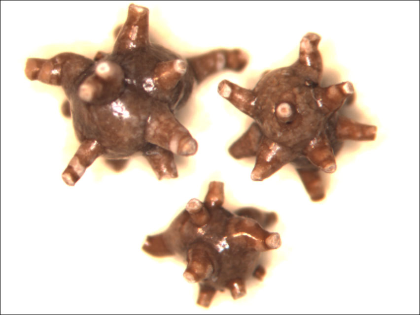
Macroscopic and Microscopic Examination
The initial step in analysis is examination of the intact calculus before fracturing so that crystals, amorphous material, or foreign bodies adhering to or embedded in the surface can be observed. The objective of fracturing is to make visible the nidus, the crystalline structure and the order of deposition of components. The true nidus is invisible because it is the first crystal or aggregate of crystals precipitated from solution and deposited at what eventually becomes the stone site. An "apparent nidus" is either a region from which crystalline forms radiate or the geometric center surrounded by concentric laminations. The apparent nidus may be roughly in the center of the stone, or it may be closer to one pole, as in a staghorn calculus. In calculi of 100 mg. or more, the apparent nidus may be visible to the unaided eye; however, in smaller stones, a dissecting microscope is required for visualization. Following fracturing of the calculus, the order of deposition of components is determined including identification of an apparent nidus, or nidi, if present, and identification of other patterns whether homogeneous, or characterized by layered, concentric or radial deposition structure.
Nidi may form from precipitation of crystals from supersaturated urine, from microscopic debris in urine, from artifacts or foreign bodies, from drugs or drug metabolites, or from calcium plaques in the renal papillae. Finding any of these components may give a clue to the pathogenesis of the stone.
In the study of multiphase stones, each distinct phase may be separated for analysis. Some components are sufficiently characteristic in appearance as to obviate further analysis, but incongruities in consistency, density, or pigmentation may indicate the presence of interstitial components, which need further elucidation by integrative methods.
X-ray diffraction identifies the constituents of a calculus by their unique diffraction patterns or "fingerprints" produced by monochromatic x-ray bombardment of crystalline material. The x-rays, when traveling intramolecular distances, are diffracted or reflected in characteristic patterns related to the structure of the crystals. The reflected x-rays may be used to produce a diffractogram composed of peaks or "maxima" registered as the sample rotates through a succession of angles. This allows definite identification of an unknown crystalline substance.
The major advantage of x-ray diffraction is its almost absolute identification of crystalline materials and mixtures of crystalline materials. When amorphous and crystalline substances are mixed, other methods may confuse the two (as in the case of phosphatic calculi); x-ray diffraction selectively identifies the crystalline components. Common crystalline components of urinary calculi are all readily identifiable and potentially measurable by this method. Crystalline apatites may be identified but usually yield poor diffraction patterns because they are in a microcrystalline state and give broad, weak, diffraction bands.
The main disadvantage of x-ray diffraction is it's poor ability to identify some metabolites or amorphous materials and constituents present in only minor or trace amounts.
Infrared spectroscopy is specific, rapid, and versatile and can be used with specimens of various sizes. It uses a spectrophotometer which exposes sample molecules to infrared light. In the infrared region of the electromagnetic spectrum, light is absorbed as the vibrational stretching and bending of groups of covalently bonded atoms occur in response to excitation at specific wavelengths. Most organic and inorganic solids have absorption patterns that include several absorptance maxima at wavelengths characteristic of particular functional groups that make up the molecule. Correlation of specific observed absorption maxima of the unknown with our reference spectra allows positive identification of a sample.
Infrared spectroscopy is useful for the identification of noncrystalline materials, including amorphous and fatty substances. This gives it an advantage over x-ray diffraction, which is useful primarily for analyzing crystalline compounds. Such compounds as carbonate apatite and hydroxyl apatite, may give weak, diffuse lines on an x-ray diffraction pattern but may be identified and measured by infrared spectroscopy.
A particular application of this method is the identification of drugs and drug metabolites in urinary calculi, which often have well resolved absorption maxima. Since drugs are often partially metabolized, positive identification may depend on analytic studies, the patient's drug history, and pharmacokinetic information.
Infrared spectroscopy is useful also for the identification of the many artifacts that appear in calculi. Usually, infrared analysis will reveal the true nature of these artifacts. Other common artifacts easily identified by infrared spectroscopy are quartz and kaolin, both of which have highly characteristic spectra.
Chromatography is based on differences in the distribution ratios of the components of mixtures between a mutually immiscible mobile and a fixed phase. Isolated bands or spots are mechanically separated and then may be further analyzed. The mobile phase used in our laboratory is a combination of organic solvents, and the stationary phase is a solid. The use of thin layer (or film) silica gel chromatography in this laboratory has enabled us to separate clearly as many as five drug metabolites (and the intact drug) with long wavelength ultraviolet light to visualize each component. This analytic tool is most helpful when the weight percentage of a drug or other organic component of the calculus is very small. Microgram quantities of each component are readily separated and easily discernible. In many cases, a quantitative procedure can be applied to drugs and metabolites and exact quantities of "standards" used as references.
Many organic compounds exhibit fluorescence when excited by light at specific wavelengths. The excited component emits unpolarized light of a wavelength greater than that of the exciting light, owing to the return of excited electrons to a more stable position. Compounds exhibiting this property may be detected or measured at concentrations far below what is required by other analytic methods.
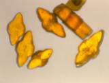
Polarization Optical Crystallography
With a polarizing microscope it is possible to establish the presence of crystalline material and obtain optical constants that are helpful in its identification; the polarizing microscope also may be the only available means for observing and identifying crystals. Some optical properties that may be determined are the crystal system (monoclinic, hexagonal, tetragonal, rhombic, orthorhombic, etc.), the optical sign, the refractive index, the angle of extinction, and the presence of birefringence. These properties may be compared with known data. If sufficient material is present and there is doubt about the identification, confirmatory studies by other methods are necessary.
It should be remembered that proper use of the polarizing microscope requires a great deal of skill, patience and experience. It cannot be used on a casual basis. Polarizing microscopy is not applicable to analysis of calculi that contain only amorphous compounds, such as large phosphatic calculi, or those consisting of complex salts of uric acid.
Microchemical tests play an important supportive role in the analysis of renal calculi and often indicate a need for confirmatory tests by other methods. There is good correlation between commonly used chemical tests and x-ray diffraction, but it must be remembered that chemical tests are specific only for particular elements or molecular functional groups and are subject to false-positive or false-negative reactions. Results must be interpreted in conjunction with other compatible criteria. It also is important to understand that attempts at measurement (as opposed to identification) by these methods are fruitless and should not be relied upon because the sensitivity of the reagents to individual constituents varies greatly and may be misleading. Microchemical tests are most helpful in analyzing amorphous or some crystalline material but are of little or no value in analyzing drug metabolites or artifacts.
Sample size is often the factor that limits the application of these tests because they are performed by using a small amount of powdered specimen combined with the appropriate reagents on a porcelain spot plate or on a microscope slide. We find that in small samples the best results are obtained with immediate nondestructive methods, such as infrared spectroscopy; if chemical tests are needed, they are then performed on the specimen extracted from the infrared sample pellet.
Ultraviolet-Visible Spectroscopy
Ultraviolet spectrophotometry is expedient and specific for the determination of drugs and/or drug metabolites in kidney stones. Its desirability is its ability to detect and identify components of calculi which are soluble in water, alcohols, dilute mineral acids and other solvents. The solubilized compounds can be detected and identified with concentrations as low as micrograms per milliliter, making it one of the most sensitive of all spectroscopic procedures for the analysis of calculi.
Photomicrography is available for a small additional charge on a routine, but optional basis. This service provides two (2) photographs of the specimen; one of the exterior and another of the interior, when possible.